Malignant Melanoma
Expert care for melanoma, the most serious type of skin cancer, with advanced treatment options.
Malignant Melanoma
Solid Tumors
Overview
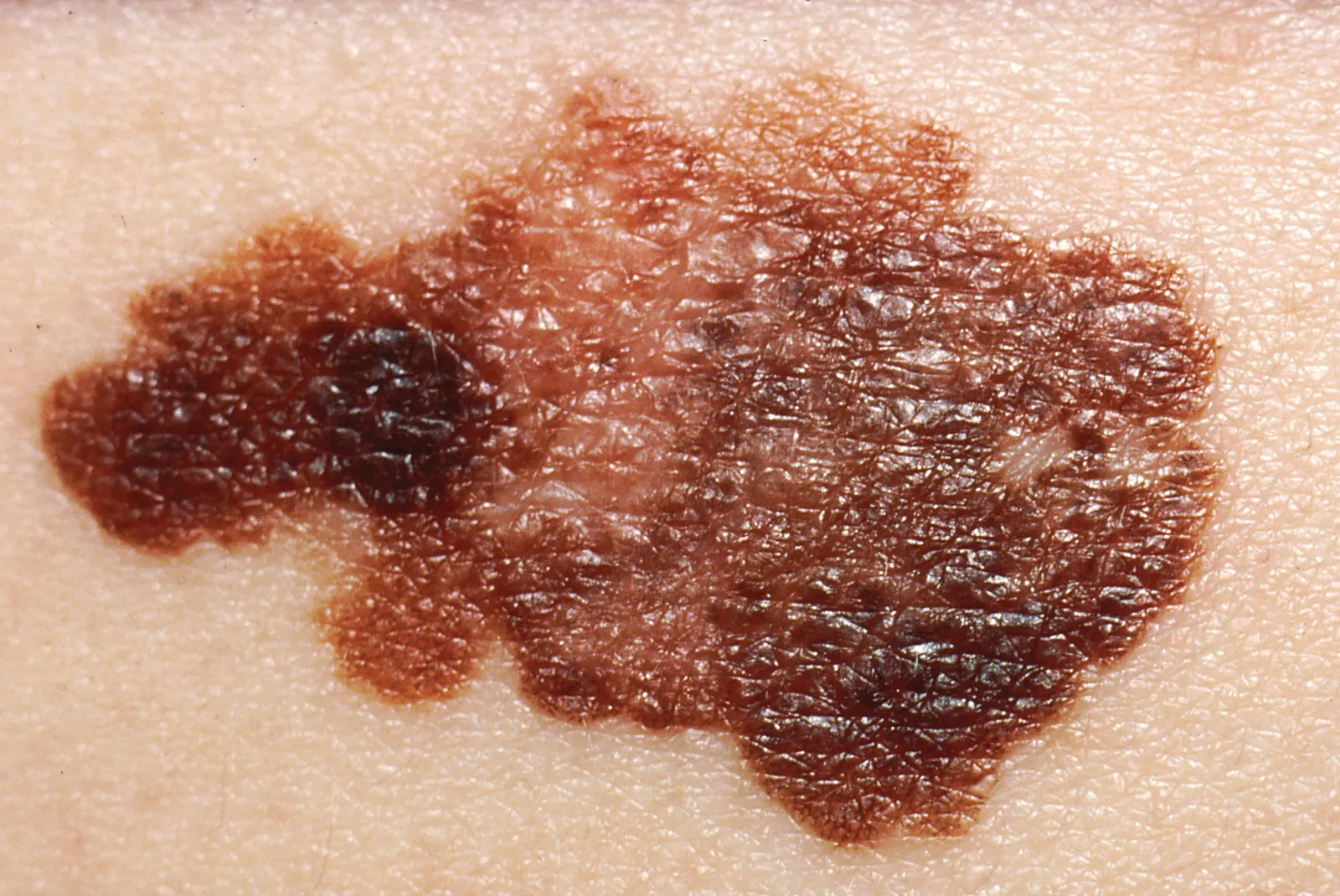
Malignant Melanoma is the most aggressive and lethal form of skin cancer, originating in the melanocytes—the pigment-producing cells located in the basal layer of the epidermis that give skin its color. While melanoma accounts for only about 1% of all skin cancers, it is responsible for the vast majority of skin cancer deaths due to its high propensity to metastasize early and rapidly. The primary driver of melanoma is exposure to ultraviolet (UV) radiation from sunlight or tanning beds, which causes DNA damage and mutations in skin cells.
Risk factors include a history of severe, blistering sunburns, fair skin that burns easily, light hair and eyes, a high number of moles (>50), presence of atypical moles (dysplastic nevi), family history of melanoma, personal history of skin cancer, and immunosuppression. Melanomas can develop anywhere on the skin, including non-sun-exposed areas, and can also occur in the eyes (uveal melanoma) or mucosal surfaces. The classic clinical tool for identifying potential melanoma is the ABCDE criteria: Asymmetry, Border irregularity, Color variation, Diameter > 6 mm, and Evolving (changing in size, shape, color, or symptoms like itching and bleeding).
Melanoma grows horizontally within the epidermis (radial growth phase) before invading vertically into the deeper dermis (vertical growth phase), where it gains access to lymphatic and blood vessels, leading to spread to regional lymph nodes, lungs, liver, brain, and bones.
When to Consult
If you have a suspicious mole (changing size, shape, color), new pigmented lesion, or confirmed melanoma diagnosis.
What to Bring
Biopsy reports, photographs of the lesion, sentinel lymph node biopsy results, BRAF mutation testing, CT/MRI scans if advanced, and any previous treatment records.
Risk Factors
Causes
Treatment Options
Wide Local Surgical Excision
Wide local excision is the primary curative treatment for localized melanoma (Stages I-II). Under local anesthesia, the surgeon removes the biopsy site along with a predefined margin of surrounding healthy skin and subcutaneous fat down to the deep fascia. The surgical margins are strictly determined by the Breslow thickness of the primary tumor: Melanoma in situ requires a 0.5 cm margin; tumors <= 1.0 mm thick require a 1.0 cm margin; tumors 1.01 to 2.0 mm thick require a 1-2 cm margin; and tumors > 2.0 mm thick require a 2.0 cm margin. Wide margins ensure that any microscopic satellite tumors in the surrounding skin are removed, minimizing the risk of local recurrence.
Sentinel Lymph Node Biopsy and Lymphadenectomy
Sentinel Lymph Node Biopsy (SLNB) is a staging procedure performed to identify micro-metastases in regional lymph nodes. A radioactive tracer and blue dye are injected at the primary tumor site, and a handheld gamma probe is used to locate the first lymph node(s) that drain the tumor (the sentinel node). This node is surgically removed and examined. If the sentinel node is negative, no further lymph node surgery is needed. If the sentinel node is positive, it indicates lymphatic spread. Historically, a complete lymph node dissection (removing all nodes in the groin, axilla, or neck) was performed, but current guidelines prefer active surveillance with regular ultrasounds, reserving surgery for clinically palpable nodes.
Adjuvant and Advanced Cancer Immunotherapy
Immunotherapy has revolutionized the management of melanoma, utilizing immune checkpoint inhibitors to activate the body's immune system against cancer cells. For patients with high-risk Stage II or III melanoma after complete surgical resection, adjuvant immunotherapy with Pembrolizumab or Nivolumab (anti-PD-1 antibodies) for one year significantly reduces recurrence risk. In the advanced or metastatic (Stage IV) setting, doublet immunotherapy combining Nivolumab with Ipilimumab (anti-CTLA-4) is the first-line standard of care. This combination can achieve deep, durable responses and long-term survival in a significant proportion of patients, though it carries a high risk of immune-related side effects.
BRAF and MEK Targeted Therapies
Approximately 40-50% of cutaneous melanomas harbor a mutation in the BRAF gene, most commonly the BRAF V600E mutation, which leads to constitutive activation of the MAPK pathway and rapid cell growth. For patients with BRAF-mutated advanced or metastatic melanoma, targeted therapy combining a BRAF inhibitor with a MEK inhibitor is highly effective. Standard oral combinations include Dabrafenib plus Trametinib, Vemurafenib plus Cobimetinib, or Encorafenib plus Binimetinib. These targeted therapies achieve rapid tumor shrinkage and symptom relief, and are also approved for use in the adjuvant setting for Stage III BRAF-mutated melanoma to prevent recurrence.
Frequently Asked Questions
Q. What is the most effective treatment for Malignant Melanoma?
The most effective treatment for Malignant Melanoma depends on the stage, location, molecular profile of the tumor, and the patient's overall health. Dr. R. Srinath Bharadwaj provides personalized protocols including chemotherapy , immunotherapy , targeted therapy , or combination approaches.
Q. Where can I get expert treatment for Malignant Melanoma in Hyderabad?
You can consult Dr. R. Srinath Bharadwaj, a leading Medical Oncologist, at the American Oncology Institute, Nallagandla, Hyderabad. Call +91 91213 36638 to schedule an appointment.
Q. What documents should I bring for a Malignant Melanoma consultation?
Please bring all recent biopsy reports, imaging scans (CT, MRI, or PET-CT), tumor markers, blood test results, and any previous treatment or surgery details to help outline your care plan.